
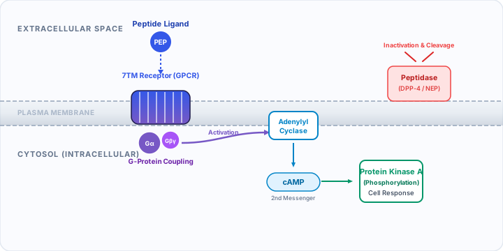
1. The Lock-and-Key Signaling Model
In human physiology, biological regulation depends on constant intercellular communication. Because cells are separated by extracellular fluids and membranes, they require specialized signaling molecules to transmit instructions. Peptides function as the primary precision messengers of this system [1].
The classical conceptual model for peptide action is the lock-and-key model, first proposed by Emil Fischer and refined into modern induced-fit conformational theory. The cell-surface receptor acts as the lock, presenting a stereospecific binding pocket configured with complementary electrical charges, hydrophobic surfaces, and hydrogen-bonding sites. The peptide acts as the flexible key, docking into the receptor pocket and inducing a structural rearrangement that sets off internal biochemical activity [2].
2. Cell-Surface Receptors: Why Peptides Stay Outside
The outer boundary of every human cell is a phospholipid bilayer, a dense, hydrophobic barrier that prevents the uncontrolled passage of water-soluble molecules. Because peptides are polar, hydrophilic chains with charged N- and C-termini and diverse side chains, they cannot passively diffuse across cell membranes like lipophilic steroid hormones do [3].
Consequently, peptides do not enter the cytoplasm to exert their primary biological effects. Instead, they interact exclusively with transmembrane receptors whose extracellular domains face the external fluid and whose intracellular domains extend into the cytosol. By spanning the plasma membrane, these proteins convert an external chemical binding event into an internal biochemical signal, a process known as signal transduction [1].
3. GPCRs: The Primary Peptide Target
More than 70% of known endogenous peptide hormones and commercial peptide drugs interact with G protein-coupled receptors (GPCRs), specifically members of the Class A (rhodopsin-like) and Class B (secretin-like) subfamilies [4].
In plain language: GPCRs are proteins on the cell surface that receive messages. When a matching molecule binds, the receptor can activate a G protein inside the cell, starting a chain of signals. Receptor activation explains a possible biological effect; it does not by itself prove a clinical benefit. Read the NCBI cell-signaling explanation.
GPCRs possess a conserved structural architecture composed of seven hydrophobic alpha-helices spanning the lipid membrane (7TM architecture), connected by three extracellular and three intracellular loops. When a peptide docks into the extracellular binding groove:
- The receptor undergoes an outward movement of transmembrane helices 5 and 6, opening an intracellular cavity [2].
- This conformational change recruits a heterotrimeric G-protein complex (composed of Gα, Gβ, and Gγ subunits) to the intracellular face of the receptor.
- The Gα subunit releases guanosine diphosphate (GDP) and binds guanosine triphosphate (GTP), causing it to dissociate from the Gβγ dimer and engage downstream effector enzymes [4].
Robert Lefkowitz and Brian Kobilka were awarded the 2012 Nobel Prize in Chemistry for elucidating the precise molecular mechanisms and crystal structures of these GPCR signaling complexes [2].
4. Second Messenger Cascades & Protein Kinases
Once activated, the Gα subunit does not perform the end physiological action directly; rather, it activates enzymatic amplifiers that generate thousands of small intracellular molecules called second messengers [1]:
- Gαs (Stimulatory Pathway): Activates adenylyl cyclase, an enzyme that converts ATP into cyclic adenosine monophosphate (cAMP). cAMP then binds and activates Protein Kinase A (PKA), which phosphorylates metabolic enzymes and nuclear transcription factors like CREB. Example: Glucagon and GLP-1 receptor activation [5].
- Gαi/o (Inhibitory Pathway): Inhibits adenylyl cyclase, lowering intracellular cAMP and closing voltage-gated calcium channels. Example: Somatostatin and opioid receptor activation.
- Gαq/11 Pathway: Activates phospholipase C beta (PLCβ), cleaving membrane PIP₂ into inositol 1,4,5-trisphosphate (IP3) and diacylglycerol (DAG). IP3 releases calcium from the endoplasmic reticulum, while DAG activates Protein Kinase C (PKC). Example: Angiotensin II and Oxytocin receptor activation [3].
This enzymatic cascade acts as an exponential signal amplifier: a single peptide molecule binding to one cell-surface receptor can generate millions of phosphorylated enzymes inside the cell within milliseconds.
5. Agonists, Antagonists & Biased Signaling
Depending on their chemical structure, synthetic peptide therapeutics can interact with receptors in several pharmacologically distinct ways [6]:
Full Agonists: Replicate or exceed the maximal biological response of the natural endogenous peptide (e.g., semaglutide acting on the GLP-1 receptor).
Partial Agonists: Bind the receptor but elicit a sub-maximal biological response even at saturating concentrations, acting as functional buffers that can compete with native hormones.
Antagonists: Occupy the orthosteric binding site without activating the receptor, blocking endogenous signaling. For example, cetrorelix and ganirelix are peptide GnRH antagonists used in reproductive medicine to prevent premature ovulation surges [6].
Biased Ligands: Selectively stabilize conformations that activate canonical G-protein cascades while minimizing beta-arrestin recruitment, potentially reducing receptor desensitization and side effects.
6. Endocrine, Paracrine & Autocrine Signaling
Peptide signaling occurs across three primary anatomical ranges, depending on where the peptide is secreted and how far it travels [1]:
- Endocrine Signaling: Peptides are secreted into the bloodstream by specialized neuroendocrine cells or glands (e.g., insulin from the pancreas, vasopressin from the posterior pituitary) and travel systemically to target distant organs.
- Paracrine Signaling: Peptides are released into the local interstitial fluid to act on neighboring cells within the same tissue, without requiring systemic circulation (e.g., somatostatin delta cells regulating adjacent alpha and beta cells in pancreatic islets).
- Autocrine Signaling: The secreted peptide binds receptors located on the surface of the very cell that produced it, providing an immediate negative or positive autocrine feedback loop.
7. Enzymatic Breakdown & Half-Life
Because peptides coordinate dynamic, time-sensitive physiological events, the human body has evolved aggressive enzymatic mechanisms to rapidly terminate peptide signals [7].
Ubiquitous cell-surface and soluble enzymes called peptidases and proteases recognize and cleave peptide bonds within seconds or minutes:
- Dipeptidyl Peptidase-4 (DPP-4): A serine protease expressed on endothelial cells and lymphocytes that cleaves the N-terminal dipeptide from GLP-1 and GIP, rendering native GLP-1 inactive within 1.5–2 minutes in circulation [5].
- Neutral Endopeptidase (NEP / Neprilysin): A zinc-dependent metalloprotease that cleaves natriuretic peptides, bradykinin, and enkephalins.
- Renal Filtration: Peptides smaller than ~30 kDa that escape enzymatic degradation are rapidly filtered by the glomeruli in the kidneys and degraded by tubular lysosomes.
Extending peptide half-life from minutes to days is the foundational challenge of modern peptide drug design. Strategies such as fatty acid acylation (lipidation), albumin binding, and amino acid substitution (e.g., replacing L-Ala with Aib in semaglutide) protect the molecule from peptidase access [7]. Learn more in our Metabolic Peptides Research Review.
8. Why Mechanisms Differ Between Peptides
A frequent error in public health discourse is assuming that "all peptides work alike." In reality, the mechanism of any specific peptide is defined by three strict biological parameters:
- The Identity of Its Target Receptor: A peptide designed for the melanocortin receptor (MC4R) will have no cross-reactivity with insulin or growth hormone secretagogue receptors.
- Tissue Expression Profile: If an organ does not express the matching receptor, the peptide cannot exert direct biological effects on that organ, regardless of dose.
- Downstream Effector Coupling: The intracellular G-protein or kinase network linked to the receptor determines whether the cellular response is contraction, secretion, gene transcription, or apoptosis [4].
To understand the full spectrum of structural designs and functional classes, read our companion guide on Types of Peptides.
Core Scientific Definitions
Precise biomedical definitions for terms used throughout this guide.
- Transmembrane Receptor
- A specialized protein embedded across the cell plasma membrane that binds extracellular ligands and transmits biochemical signals across the lipid bilayer into the interior of the cell.
- G Protein-Coupled Receptor (GPCR)
- A family of proteins on the cell surface that receive signals and pass messages inside the cell through G proteins. In technical terms, each receptor crosses the cell membrane seven times and can activate intracellular signaling pathways.
- Agonist
- A chemical molecule that binds to a receptor and stabilizes its active conformation, triggering a biological response.
- Antagonist
- A molecule that binds to a receptor without activating it, physically preventing or attenuating the binding of endogenous agonists.
- Circulating Half-Life (t½)
- The time required for the biological concentration of a peptide in the bloodstream to decrease by 50% through enzymatic degradation and excretion.
- Peptidase
- An enzyme that catalyzes the hydrolytic cleavage of peptide bonds, terminating peptide signaling and recycling amino acids.
Peer-Reviewed References & Primary Sources
This educational guide is built strictly upon peer-reviewed literature, official regulatory guidance, and authoritative biochemical repositories.
-
Signal Transduction (3rd Edition) Academic Press / Elsevier · 2016 · Chapters 2 & 5: G-protein Coupled Receptors and Second Messenger Cascades
-
A brief history of G-protein coupled receptors (Nobel Lecture) Nature Reviews Drug Discovery · 2013 · 12(9): 647–651
-
Molecular Cell Biology (9th Edition) W. H. Freeman · 2021 · Chapter 15: Signaling at the Cell Surface
-
G protein-coupled receptors as targets for peptide hormones Nature Reviews Molecular Cell Biology · 2002 · 3(9): 639–650
-
The Incretin System: glucagon-like peptide-1, glucose-dependent insulinotropic polypeptide, and dipeptidyl peptidase-4 Lancet · 2006 · 368(9548): 1696–1705
-
Designing peptide receptor agonists and antagonists Nature Reviews Drug Discovery · 2002 · 1(11): 847–858
-
Peptide therapeutics: current status and future directions Drug Discovery Today · 2015 · 20(1): 122–128
Educational Disclaimer: This guide is produced strictly for biomedical educational purposes and does not constitute medical advice, diagnosis, or treatment recommendations. Experimental peptides discussed herein may lack regulatory authorization for human clinical use. Consult a licensed medical practitioner before considering any therapeutic intervention.