
What biased agonism means
Biased agonism is when a drug activates a receptor in a way that favors some of that receptor's signaling pathways over others, compared with a reference molecule. The idea is also called functional selectivity or ligand bias. It applies above all to G protein-coupled receptors (GPCRs), the large family of cell-surface receptors used by many peptide hormones, including GLP-1, GIP, glucagon, ghrelin, oxytocin and the melanocortins [1][2].
The older picture treated a receptor as a simple on-off switch: a drug either turned it on (an agonist), partly on (a partial agonist), or blocked it (an antagonist). Research since the early 2000s showed that a single receptor can adopt several active shapes, and different ligands can stabilize different shapes. Each shape may couple more readily to some signaling partners than others, so two molecules that both "activate" a receptor need not produce the same mixture of signals inside the cell [2][3].
Bias is always relative. A molecule is described as biased compared with a reference, usually the body's own hormone, measured in the same cells with the same assays. Without a stated reference and system, the word has no fixed meaning. For background on receptor families, see peptide receptors and peptide signaling.
G proteins and β-arrestins: two branches of one receptor
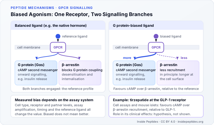
When a hormone binds a GPCR, the receptor changes shape and activates a G protein inside the cell. For receptors such as the GLP-1 receptor, the main G protein (Gαs) switches on an enzyme that produces cyclic AMP (cAMP), a small "second messenger" molecule that carries the signal onward. In pancreatic beta cells, for example, GLP-1 receptor cAMP signaling is linked to glucose-dependent insulin release.
Activated receptors are then tagged with phosphate groups by enzymes called GPCR kinases. The tagged receptor attracts β-arrestins. These adaptor proteins were first understood as "off" signals because they physically block further G protein coupling. Later work showed that β-arrestins can also organize signaling complexes of their own, influencing processes such as cell movement and survival [3].
Because G protein and β-arrestin engagement rely on partly different receptor shapes, a ligand can favor one branch. A G protein-biased agonist drives cAMP (or another G protein output) relatively more than β-arrestin recruitment; a β-arrestin-biased agonist does the reverse [2][4]. The G protein versus β-arrestin split is the most-studied example, but bias can also exist between different G protein types or between other measured outputs.
Desensitization and internalization
β-arrestins matter for how long a signal lasts. Desensitization is the fading of a cell's response during repeated or continuous stimulation. Short-term desensitization occurs over minutes and is mainly driven by β-arrestins stopping G protein coupling. Longer-term desensitization, called downregulation, develops over hours to days and involves pulling receptors into the cell, breaking some down in lysosomes and making fewer new receptors [5].
Internalization is the step in which activated receptors are drawn inside the cell in small membrane bubbles. From there, a receptor may be recycled to the surface or degraded. Some receptors continue signaling from inside the cell, so internalization does not simply equal "off" [4].
This is why biased agonism attracts interest in drug design. In principle, a ligand that recruits less β-arrestin might leave more receptors at the cell surface for longer, changing how a response holds up over time. In mouse and cell studies of experimental GLP-1 receptor agonists, compounds that kept the receptor at the cell surface produced greater long-term insulin release, an effect that depended on reduced β-arrestin recruitment and faster drug unbinding [6]. These were laboratory compounds, not approved medicines.
How bias is measured
Measuring bias requires more than comparing two numbers. Researchers typically:
- Choose a reference ligand, usually the native hormone.
- Measure full concentration-response curves for each ligand in at least two pathway assays, such as cAMP production and β-arrestin recruitment, in the same cell system.
- Fit the curves to a pharmacological model that separates how tightly a ligand binds (affinity) from how effectively it activates each pathway (efficacy).
- Compare each ligand's pathway preference with the reference ligand's preference.
A widely used approach, based on the Black and Leff operational model, summarizes each ligand-pathway pair as a "transduction coefficient", written log(τ/KA). Subtracting the reference ligand's value and then comparing pathways gives a relative bias estimate with a confidence interval. The method was designed so that the estimate is less sensitive to how many receptors the test cells carry [7][1].
Results are often reported as a "bias factor". This is a ratio of ratios: it states how much more one pathway is favored than the reference ligand favors it, under the conditions tested. It is not a fixed physical property of the molecule.
Why measured bias depends on the assay system
A hallmark of functional selectivity is that it depends on the cell type. That makes it hard to carry results from engineered test cells into the tissue where a drug actually acts [7]. Several factors can change the answer:
- System bias. Cells differ in receptor numbers and in the levels of G proteins, kinases and β-arrestins. A cell rich in one partner can make every ligand look as if it favors that pathway [2].
- Observation bias. Assays amplify signals differently. cAMP assays are highly amplified, so even weak activation can produce a full response, while β-arrestin recruitment assays are often less amplified. A low-efficacy drug can therefore appear "G protein-biased" simply because the β-arrestin assay cannot detect its weaker activity [8].
- Timing and location. Pathways rise and fall at different rates, and receptors can keep signaling after internalization, so a reading taken at one time point captures only part of a ligand's profile [4].
- Choice of reference. Changing the reference ligand changes the bias value of every other ligand.
The opioid field shows why this matters. Several μ-opioid receptor drugs were developed as G protein-biased agonists, with the hypothesis that less β-arrestin activity would mean fewer side effects. A 2020 study using assays designed to avoid limited signal windows found that oliceridine, PZM21 and SR-17018 had low intrinsic efficacy across pathways, and proposed low efficacy as a possible alternative explanation for their improved side-effect profile in animal experiments [8]. A reported bias, in other words, may sometimes reflect partial agonism rather than true pathway selectivity.
Peptide example: tirzepatide at the GLP-1 receptor
Tirzepatide is a single peptide that activates both the GIP receptor and the GLP-1 receptor. A 2020 paper in JCI Insight, written largely by scientists at its manufacturer, reported three main pharmacological findings [9]:
- Imbalance. Based on calculated receptor occupancy at clinically used exposures, tirzepatide engages the GIP receptor to a greater degree than the GLP-1 receptor.
- Bias at the GLP-1 receptor. In cell-based assays, tirzepatide mimicked native GIP at the GIP receptor but, at the GLP-1 receptor, favored cAMP generation over β-arrestin recruitment compared with native GLP-1. It also caused weaker GLP-1 receptor internalization.
- An islet experiment. In primary mouse islets, β-arrestin 1 limited the insulin response to GLP-1 but not to GIP or tirzepatide. The authors suggested that this biased agonism may enhance insulin secretion.
An independent 2021 study using different techniques reported that tirzepatide and another dual agonist preserved maximal cAMP production despite only partial recruitment of the G protein, together with reduced receptor internalization at both receptors, relative to native and acylated GLP-1 agonists including semaglutide [10]. These findings broadly agree that tirzepatide's GLP-1 receptor signaling profile differs from that of GLP-1, though they come from cell systems.
What is shown and what is still a hypothesis
| Claim | Status |
|---|---|
| Tirzepatide favors cAMP over β-arrestin recruitment at the GLP-1 receptor, relative to GLP-1 | Reported in cell assays by more than one group [9][10] |
| Tirzepatide drives less GLP-1 receptor internalization than GLP-1 | Reported in cell systems [9][10] |
| β-arrestin 1 limits GLP-1's insulin response but not tirzepatide's | Shown in mouse islets [9] |
| Bias explains tirzepatide's weight-loss or glucose results in people | Hypothesis; not tested directly in humans |
| Biased drugs generally have fewer side effects | Not established; some apparent bias reflects low efficacy [8] |
Clinical trials of tirzepatide measure outcomes such as HbA1c and body weight. They do not separate the contribution of GLP-1 receptor bias from that of GIP receptor activity, the drug's long half-life, or its overall exposure. Native GLP-1 itself is cleared within minutes, so it is not a direct clinical comparator for any long-acting medicine. Reviews of the field describe "designer" biased agonists as a prospect that still requires links between laboratory profiles, structure and outcomes [4][2].
A careful summary for readers is: laboratory studies report that tirzepatide is a biased agonist at the GLP-1 receptor; whether this bias contributes to its clinical effects in people is an open question. For how to weigh cell, animal and human evidence, see why animal studies don't always predict human results.
Core Scientific Definitions
Precise biomedical definitions for terms used throughout this guide.
- Biased agonism (functional selectivity)
- The ability of a ligand to activate some of a receptor's signaling pathways more than others, measured relative to a reference ligand acting on the same receptor in the same system.
- β-arrestin
- One of two intracellular adaptor proteins (β-arrestin 1 and 2) that bind activated, phosphorylated G protein-coupled receptors, block further G protein coupling, help move receptors into the cell and can scaffold their own signaling complexes.
- Receptor desensitization
- A decline in a cell's response to repeated or continuous stimulation of a receptor, occurring over minutes (acute) or, as downregulation, over hours to days.
- Receptor internalization
- The movement of a cell-surface receptor into the cell inside membrane vesicles after activation, after which it may be recycled to the surface or degraded.
- System bias
- Apparent pathway preference that comes from the cell or tissue being studied, such as how many receptors or signaling proteins it contains, rather than from the ligand itself.
Peer-Reviewed References & Primary Sources
This educational guide is built strictly upon peer-reviewed literature, official regulatory guidance, and authoritative biochemical repositories.
-
Signalling bias in new drug discovery: detection, quantification and therapeutic impact Nature Reviews Drug Discovery · 2013
-
Biased signalling: from simple switches to allosteric microprocessors Nature Reviews Drug Discovery · 2018
-
Transduction of receptor signals by beta-arrestins Science · 2005
-
Mechanisms of signalling and biased agonism in G protein-coupled receptors Nature Reviews Molecular Cell Biology · 2018
-
GPCR desensitization: Acute and prolonged phases Cellular Signalling · 2018
-
Targeting GLP-1 receptor trafficking to improve agonist efficacy Nature Communications · 2018
-
A simple method for quantifying functional selectivity and agonist bias ACS Chemical Neuroscience · 2012
-
Low intrinsic efficacy for G protein activation can explain the improved side effect profiles of new opioid agonists Science Signaling · 2020
-
Tirzepatide is an imbalanced and biased dual GIP and GLP-1 receptor agonist JCI Insight · 2020
-
Spatiotemporal GLP-1 and GIP receptor signaling and trafficking/recycling dynamics induced by selected receptor mono- and dual-agonists Molecular Metabolism · 2021
Educational Disclaimer: This guide is produced strictly for biomedical educational purposes and does not constitute medical advice, diagnosis, or treatment recommendations. Experimental peptides discussed herein may lack regulatory authorization for human clinical use. Consult a licensed medical practitioner before considering any therapeutic intervention.